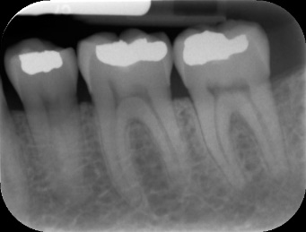
The procedure is very simple. This is an intraoral type of X-ray, where the film is inside the mouth. The X-ray technician will give you all the instruction, place the film inside the mouth and adjust the tube. You have to take off all metal things you have on you before the procedure including jewelry.
Uses:
The main use is to diagnose conditions of teeth and the surrounding tissues. Sometimes dentists are not sure whether there is a periapical process, so they have to use this X-ray to set up a diagnosis. Other times, they use it as a part of the treatment, for example, when they need to see dental caries and its depth. When it comes to decay, dentists can monitor which tissues it has affected, which surfaces, and whether there is some proximity to the dental pulp. This is all very useful when removing the decay and placing the filling. For periapical conditions, an x-ray can show if the infection has spread from the pulp to the surrounding bone, whether it is a localized infection or it has spread. It helps with diagnosing fractures of the teeth and the bone, cysts, tumors, the number of roots, number of canals, the location of the apex and more.
When dentists plan on doing a root canal procedure, they need all the information about the number and position of roots and canals. If they don’t use a microscope, the visibility is limited, so periapical X-rays provide a detailed picture of both the crown and the roots. Once the therapy is done, dentists can request for another periapical X-ray after a certain time, to get an insight of the success of the therapy.
Most of the impacted teeth are easily diagnosed with this type of X-ray. Dentists are able to see their position, the position compared to the surrounding tissues, the thickness of the bone, the proximity to nerves and more. The same thing applies for extractions. The roots of the upper molars can be close to the walls of the sinus, so the periapical image can show that. 3D scans are much more detailed and provide a better picture, but dentists still use the periapical ones in some cases.

