


What is Leukoplakia?
 The term Leukoplakia doesnt describe a specific disease but a clinical observation. Clinically Leukoplakia looks like a white patch which cannot be removed. It can have different surfaces such as smooth, fissured, varicous or speckled. Risk factors of Leukoplakia are the “6 s”
– smoking
– spirit
– sharp tooth( traumatic origin)
– spicy food
– syphilis
– sepsis( specifically candida infection and human papilloma virus).
Beside the etiological factors above a Vitamin A deficiency can cause a Leukoplakia as well.
Leukoplakia can be differentiated in 4 clinical stages based on its size an 5 stages based on its histopathological status which are:
– no dysplasia
– mild dysplasia
– moderate dysplasia
– severe dysplasia(with high risk to be transformed to oral cancer)
– squamous cell carcinoma in situ with a poor prognosis and high letality

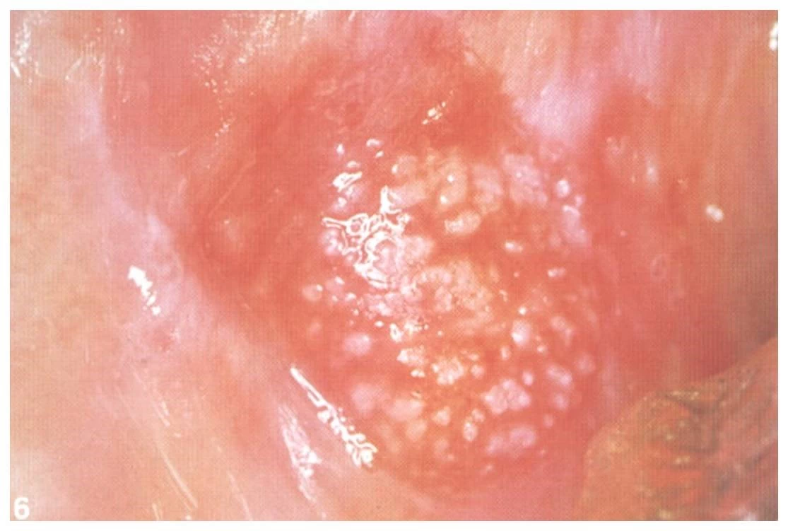
The surface of Leukoplakia can vary not only in its size but as well in its clinical appearance which could be :
-smooth
-fissured
-verruciform
-speckled
picture different surfaces
A biopsy would be needed in case a leukoplakia is detected as the following lesions look similar:
– White spongus nevus
– Lichen planus
– Acute pseudomembranous candida albicans
– Morsicatio buccarum
– Leukoedema
Leukoplakia can be treated by non surgical approach is:
-Vitamin A
-Vitamin E
-Vitamin C
or others.
Then the invasive approach could be performed by
-Cryotherapy
-Carbon dioxide or Erbium Yag Laset therapy(Excision or Vaporization)
-Electrocautery
-Surgical excision
Always watch out for uncommon appearances in your mouth as they can be related to serious conditions like oral cancer or other systemic diseases.